

Foetal Growth Restriction in Pregnancy: Causes, Diagnosis and Management
Sonam was 32 weeks pregnant when a routine growth scan showed that her baby was measuring smaller than expected. She felt well, the baby was moving normally, and there had been no obvious warning signs. This is often how foetal growth restriction is first identified.
A small measurement does not automatically mean something is wrong. Some babies are constitutionally small and continue to grow normally, while others are not reaching their expected growth potential because of placental, maternal, or foetal factors.
As a practising obstetrician and gynaecologist managing high-risk pregnancies, I do not assess growth from one scan value alone. I look at the growth trend, Doppler blood flow, maternal health, and the overall clinical picture before deciding whether closer monitoring is needed.
In this article, I explore what foetal growth restriction means, its common causes, how ultrasound and Doppler help confirm the diagnosis, and how careful surveillance guides delivery timing and supports safer outcomes for mother and baby.
Also Read:
Understanding Foetal Growth Restriction and Normal Growth Patterns
Growth assessment sits inside routine antenatal care because it is one of the clearest windows into how well the placenta is working. A small measurement opens a question rather than settling one.
Foetal growth restriction means a baby is not reaching its expected growth potential during pregnancy. It is identified by assessing serial growth measurements, ultrasound findings, Doppler blood flow and the wider maternal clinical picture rather than relying on one small measurement alone.
 Foetal Growth Restriction Is More Than a Small Baby
Growth restriction and small size are often treated as the same finding. Separating them is the first clinical task, and it decides how closely a pregnancy is watched.
Foetal growth restriction describes a baby whose growth has been limited below its own genetic potential. Small for gestational age says something different. It means the baby measures below a chosen percentile on a chart. A constitutionally small baby follows its own line steadily, shows normal Doppler flow, and is usually healthy. Restriction looks different. The baby drifts away from its earlier trajectory because the placenta is not supplying it well. Treating every small baby as restricted leads to deliveries that were never needed.
Foetal Growth Monitoring Assesses Growth Across Pregnancy
Assessment is a comparison over time rather than a verdict at one appointment, which is why accurate pregnancy dating matters as much as the scan itself.
Growth is plotted against validated gestational-age reference charts and interpreted alongside maternal and pregnancy factors. Accurate dating from an early scan underpins all of it. Wrong dates mean every later measurement is read against the wrong expectation. After that, I watch the shape of the line. A baby sitting on the tenth percentile at every scan behaves differently from one that has fallen from the fiftieth to the tenth. The second concerns me more.
Growth assessment is a comparison over time. The purpose is to find the baby whose growth has slowed, not simply the baby who happens to measure small at one appointment.
Maternal, Placental and Foetal Causes of Growth Restriction
Restricted growth is a finding with something behind it. Identifying that cause shapes how closely the pregnancy is monitored and how early any concern is likely to appear.
Maternal and Placental Causes
Most cases trace back to the placenta, either directly or through a maternal condition affecting placental blood supply.
- Hypertensive conditions: Chronic hypertension and pre-eclampsia reduce blood supply to the placenta.
- Placental insufficiency: The placenta transfers less oxygen and fewer nutrients than the baby needs.
- Maternal medical conditions: Chronic kidney disease, autoimmune disease and diabetes with vascular complications may contribute to impaired foetal growth.
- Nutritional and lifestyle factors: Significant maternal anaemia, poor nutrition and smoking are associated with restricted growth.
Foetal Factors and Closer Surveillance
A smaller group of cases originate with the baby rather than the placenta, and some pregnancies carry enough risk to warrant monitoring from the start.
- Chromosomal and structural conditions: Certain genetic abnormalities and congenital anomalies limit growth from early pregnancy.
- Selected infections: A small number of infections in pregnancy restrict foetal growth.
- Known risk factors: A previous growth-restricted pregnancy or maternal vascular disease prompts closer foetal growth monitoring.
Identifying a cause guides how intensively a pregnancy is watched. Where no cause is found, surveillance still continues, because the growth pattern itself directs the decisions that follow.
Diagnosis and Doppler Assessment in Foetal Growth Restriction
Diagnosis is a staged process. No single measurement confirms growth restriction. Findings build up across clinical examination, serial ultrasound and blood flow assessment before a conclusion is reasonable.
From Fundal Height to Ultrasound Confirmation
Measuring the abdomen with a tape is where screening begins, though it was never designed to carry a diagnosis alone.
Fundal height is a screening step that is quick, useful and imperfect. Maternal obesity, fibroids and multiple pregnancy reduce its reliability, so a normal tape measurement does not rule out a concern. Ultrasound then takes over. Estimated foetal weight and abdominal circumference are the primary measures, with amniotic fluid volume supporting them. One scan gives a position. Repeated scans give a direction.
 How Does Doppler Ultrasound Assess Foetal Growth Restriction?
Doppler ultrasound assesses blood flow between the placenta and baby, especially through the umbilical artery. Abnormal resistance, absent flow, or reversed end-diastolic flow can indicate placental insufficiency and help doctors judge severity, surveillance frequency, and the safest timing of delivery.
Diagnosis emerges from a pattern. Growth trend, blood flow and maternal assessment are read together, which is why I ask for a repeat scan before drawing conclusions from one report.
Monitoring, Delivery Timing and Newborn Outcomes
Management here centres on surveillance rather than treatment. The clinical question becomes how long a pregnancy can safely continue and at what point delivery becomes the safer option.
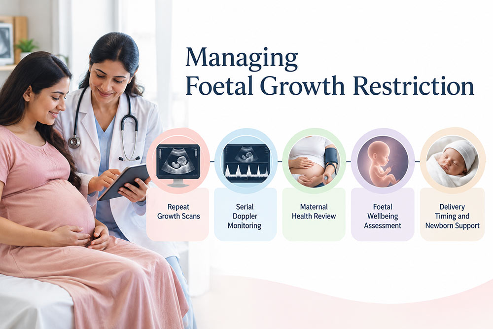
- Surveillance schedule: Repeat growth scans, serial umbilical artery Doppler and foetal wellbeing assessment, at intervals decided by how severe the findings are.
- No growth medicine: Once placental insufficiency is established, no medication reliably makes a baby grow. Careful monitoring becomes the intervention itself.
- Maternal optimisation: Managing hypertension and relevant medical conditions protects maternal health and supports safer pregnancy management.
- Delivery timing: The risks of prematurity are weighed against declining placental reserve at every review, which is the central judgement in these pregnancies.
- Newborn support: Babies born with low birth weight may need paediatric monitoring or a period of neonatal care after delivery.
- After birth: Placental examination, paediatric follow-up and a review of risk in future pregnancies.
Structured surveillance helps doctors recognise deterioration early, choose the safest time for delivery, and arrange neonatal support where it is needed. That combination is what protects outcomes here.

Also Read:
- Pregnancy After 35: Risks, Care, and Healthy Outcomes
- High-Risk Pregnancy: Causes, Risk Factors, and Safe Pregnancy Care
Conclusion
A small baby and a growth-restricted baby are assessed differently. The distinction rests on the growth trend and Doppler findings rather than any single measurement.
Where foetal growth restriction is confirmed, foetal growth monitoring and ongoing surveillance become central to care. How often the baby is monitored and when delivery is planned are important decisions that help protect both mother and baby.
A growth concern identified early and monitored appropriately can often be managed with careful clinical follow-up. Early identification helps doctors make timely decisions aimed at achieving the safest possible outcome for mother and baby.
If your scan has raised concerns about your baby’s growth, book a consultation with Dr. Madhu Goel for personalised assessment and monitoring guidance.